A HIDA (hepatobiliary iminodiacetic acid ) scan is an imaging test. It can be used to check for problems in the liver, gallbladder, and the tubes inside and outside the liver (bile ducts).
During the test, a small amount of radioactive substance (tracer) is injected into a vein in your arm or hand. Pictures are then taken to track the movement of the tracer. The test takes about 2 hours. In some cases, more pictures may need to be taken after a wait of 4 hours. You'll be told as the test progresses how long your test may take.
Before the test
- Follow any directions you're given for not eating or drinking before the procedure. Your doctor will give you instructions if required.
- Tell your doctor what medicines you're taking. This includes vitamins, herbs, and over-the-counter medicines. You may be told to stop taking some or all of them in the days before the test.
- Follow any other instructions you're given to get ready for the test.
What to tell the technologist
Let the technologist know if you:
- Are taking any medicines or have allergies to any medicines. Some medicines may prevent accurate test results.
- Had recent X-rays or tests that used barium.
- Had recent surgery or illness.
- Have other health problems, such as diabetes.
- Are pregnant or think you might be pregnant.
- Are breastfeeding.
- Smoke or use other tobacco products.
During the test
The test is done by a nuclear medicine or radiology technologist. It can be done in a hospital or test center.
- You'll lie on your back on a table. A special camera (also called a scanner) will be positioned above your belly (abdomen).
- An I.V. (intravenous) needle or I.V. line is placed into a vein in your arm or hand. The tracer is then injected through the I.V. line.
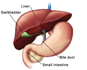
- Pictures are taken as the tracer follows the movement of bile through the liver, gallbladder, and bile ducts, and the first part of the small intestine (duodenum). Bile is a substance made by the liver that helps you digest fat.
- You'll need to lie still to help ensure that the pictures are not blurry.
- You may be given a substance by mouth or injected through a vein that causes the gallbladder to contract and release bile. Be sure to let the technologist know if you feel discomfort. This could indicate gallbladder dysfunction.
- If needed, more pictures may be taken after 4 hours.
After the test
- The technologist will let you know when the test is completed.
- The tracer will pass out of the body in your stool and urine within 24 hours. Drink plenty of fluids to help the tracer pass.
Follow-up
Your doctor will go over test results with you when they are ready. This is likely within a few days of the test.
Possible risks
Possible risks of this imaging test can include:
- Problems at the I.V. site.
- Allergic reaction to the tracer or medicine used during the test.
- Radiation exposure from the tracer.