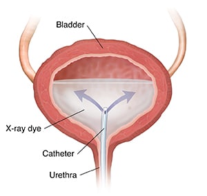
Cystography (retrograde cystography) is a detailed X-ray exam of your bladder. For this procedure, your bladder is filled with an X-ray dye (contrast medium). The dye lets your bladder be seen more clearly on the X-ray images. This procedure is done by a radiologist. This is a doctor who has special training in the use of radiation for the diagnosis and treatment of disease.
Why cystography is done
A cystography can help diagnose such bladder problems as:
- Urinary tract infections that keep coming back
- Trauma-related injuries
- Tumors
- Uncontrollable urine leakage (incontinence)
- Wounds or bursting (rupture) of your bladder wall
Getting ready for your procedure
- If instructed, empty your bowel before the exam using a medicine (laxative) or a liquid injected into your rectum (enema).
- Empty your bladder before the exam.
Tell your doctor if you:
- Have any allergies to X-ray dye.
- Take any medicines, herbs, supplements, or vitamins. This includes medicines that don't need a prescription and any street drugs you may use.
- Have any health conditions.
- Have a bladder infection.
- Are pregnant or breastfeeding, or think you may be pregnant.
- Have any history of bladder-related medical conditions.
Follow any other directions you are given to prepare for your procedure.
During your procedure
- You will change into a hospital gown and lie on an exam table. Your urethra will be numbed with an anesthetic jelly. You may also be given medicine to help you relax.
- A thin tube (catheter) will be put into your urethra up to your bladder. You will feel pressure. The dye will be slowly put through the catheter into your bladder. As your bladder fills with this liquid, you will feel the need to urinate. Tell the radiologist when this becomes uncomfortable.
- X-rays are taken of your full bladder. The catheter is removed, your bladder is then drained, and more X-rays are taken.
Possible risks
- Infection or bruising at the place where the tube is put into your urethra
- Problems due to the dye, including an allergic reaction or kidney damage
- Radiation exposure to your reproductive organs
After your procedure
- You may feel some burning when you urinate the first few times after the procedure. Drink plenty of water after the exam to help flush the dye out of your body.
- The radiologist will analyze the results and provide a report to your primary doctor (the person who referred you for the test). Your primary doctor will discuss your test results with you. They will recommend more testing or treatment as needed.
When to contact your doctor
Contact your doctor right away if any of the following occur:
- Have blood in your urine, after you have gone to the bathroom 3 times
- Have signs of infection, such as chills, fever, rapid heartbeat, or fast breathing
- Are not able to empty your bladder
Online Medical Reviewer: Neil Grossman MD
Online Medical Reviewer: Rita Sather RN
Online Medical Reviewer: Tara Novick BSN MSN
Date Last Reviewed: 09/01/2025
© 2000-2026 The StayWell Company, LLC. All rights reserved. This information is not intended as a substitute for professional medical care. Always follow your healthcare professional's instructions.